Key Takeways
Think of your body as a highly productive city where every single animal cell works as a small town. Inside the cell town, everything is organized for maximum productivity. The nucleus is like the cell's control center, making every decision. The mitochondria are like the power plants for generating energy to keep all operations running smoothly. The cell membrane functions as a security system, managing what comes in and out. Every single component has a job to do. Keep reading to learn about the major organelles of the animal cell, types of animal cells, and how they differ from the plant cell.
Introduction
Can you believe that approximately 37.2 trillion animal cells work together inside a living body, including yours? Animal cells help with energy production, breaking down food, breathing, and many other functions. All without you asking them to. That is what an eukaryotic cell does for you. But exactly what is an animal cell? And why does it matter?
An animal cell is the fundamental unit of life. From the tiniest ant to you, every creature that belongs to the animal kingdom is made of animal cells. They are eukaryotic in nature, which means they have a proper, membrane-enclosed nucleus. Unlike bacteria (which are prokaryotic and don't have a nucleus), animal cells are highly organized. Keep scrolling to learn more about its components and functions to truly understand this biology concept.
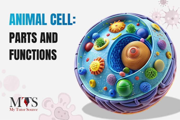
What Does an Animal Cell Actually Look Like?
If you look at an animal cell under a light microscope, you would see irregularly shaped, rounded bobs. They are often disorganized, without clear boundaries. That's not a bug, that's a feature. As they don't have a rigid cell wall, this flexibility allows your muscle cells to contract, your red blood cells to squeeze through tiny capillaries, and your nerve cells to branch out. Now let's see the animal cell organelle by organelle. Assume this cell as a cell town.
Nucleus
The nucleus is the control center of the cell. It contains DNA (your genetic blueprint) and other genetic material. From growth to protein production, the nucleus coordinates cellular activities. It's surrounded by a nuclear membrane with tiny pores.
Cell Membrane
A thin and flexible layer of lipids (fats) and proteins that surrounds the entire cell. It controls what gets in and what goes out. That is why they are semi-permeable or selectively permeable membranes.
Cytoplasm
They are jelly-like fluid that fills the cell and holds everything in its place. It works like an operating environment and a town border.
Mitrochondria
It's called the powerhouse of the cell, working as a power station. Mitochondria are responsible for:
Aerobic respiration converts glucose and oxygen into ATP (energy that the cell can actually use).
Store calcium and participate in cell signaling.
Ribosomes
Are you wondering where proteins are made in an animal cell? Ribosomes are the site of protein synthesis. They serve as tiny workers in the cell town. They read instructions from the nucleus (in the form of mRNA) and assemble amino acids into proteins. Ribosomes are found free in the cytoplasm or attached to the rough ER. It consists of 60% of RNA cytoplasmic granules and 40% proteins.
Endoplasmic Reticulum
This organelle is composed of a thin and winding network of compartments (sacs) that connect from the cytoplasm to the cell nucleus. They move the material inside the cell like a transport system. Based on its structure and function, it has two types:
Rough endoplasmic reticulum (Smooth ER): Studded with ribosomes, it mainly makes proteins.
Smooth endoplasmic reticulum (Rough ER): It is responsible for making lipids (fat) and helps detoxify chemicals.
Golgi Apparatus
Once the rough ER makes protein, it is usually not ready for use. That is where the Golgi apparatus works as a cell's post office. It receives proteins from the ER, modifies them, and packages them into vesicles. And then at last, finished products are shipped to the right place, whether inside the cell or outside it.
Lysosome
The lysosome is the solution for managing waste and recycling in a cell like a recycling center. Lysosomes contain digestive enzymes that break down old organelles, bacteria, and cellular debris.
Centrioles
Centrioles work as builders to organize cell division. When a cell is about to split, they form the spindle fibers that pull chromosomes to opposite ends of the cell.
Vacuoles
It's a fluid-filled cell organelle enclosed by a membrane. They serve as storage warehouses. They are responsible for storing food, water, carbohydrates in the form of sugars, and waste materials.
Still struggling to picture it? Watch what is inside an animal cell in 3D.
What are Kinds Of Animal Cells?
There are many different types of animal cells depending on their function and location in the body. Here are some common types:
Skin Cells
There are two main types of skin cells
Kartinocytes: produce protein.
Melanocytes: give skin its color.
Muscle Cells
Muscle cells help move an animal's limbs and organs.
Blood Cells
It has two types:
Red blood cells: Make 99% of blood cells and deliver oxygen from the lungs to the rest of the body.
White blood cells: Help organisms fight against infection and disease by killing bacteria.
Fat Cells
Fat cells store fat and other lipids as energy reserves to provide the body with energy.
From IGCSE to IB and the Cambridge biology curriculum, you are expected to draw an animal cell. But its complex structure can make you confused while drawing and labeling it correctly under exam pressure. So, what really helps?
With MTS biology tutors, you don't just explain concepts. But they also help you truly understand the structure by building a clear visualization of the structure and guiding you step-by-step through drawing it on an interactive whiteboard. Here are some tips from our expert biology tutors on how to draw an animal cell structure diagram with ease:
Step: 1
Draw a large oval shape and then add one or two small circular figures within it.
Step: 2
Draw two circles, one for the cell nucleus and the second for the tiny gap between them. Inside, draw a small shaded circle for the nucleolus. Then draw a few wavy lines inside to show chromatin.
Step: 3
Draw small oval or rod shapes for mitochondria, a stack of curved dumbbell-shaped golgi-appractus, long, folded lines for endoplamic reticulum, small circles for lysosome, and tiny straight rectangular bars for centrioles. At the end, add small circles for vacuoles and then sprinkle tiny dots all around to show ribosomes in the cytoplasm.
Conclusion
Every human being, every athlete, artist, and student reading this right now started as a single cell. And through division and coordination of trillions of organelles, that one cell becomes you. It means an animal cell is not just a microscopic structure; it's the basic unit of life. From the nucleus controlling activities to mitochondria producing energy, every organelle has its own function.
Still wondering if the animal cell has a vacuole? Want the full breakdown and know the reason behind it? MTS expert biology tutors can help you walk through it. Moreover, whether your school follows the IGCSE or the Cambridge biology syllabus, our tutors have expertise in exactly what you are looking for. Book a free trial today and start learning with ease.
FAQs
Q1: Do animal cells have a cell wall?
No, animal cells do not have a cell wall.
Q2: Do animal cells have a vacuole?
Animal cells contain temporary and small vacuoles that help store materials and remove wastes. Moreover, they are comparatively smaller than the large central vacuole found in plant cells.
Q3: Why don't animal cells have a cell wall?
Animal cells don't have a cell wall because they require flexibility for movement and specialized cellular function. At the same time, a rigid cell wall can restrict the movement.
Q4: What does an animal cell look like?
If you see it under a microscope, it would look like a round or irregular blob. It's more like a soft, jelly-like structure, you can visibly see:
Cell membrane: outer thin layer.
Cytoplasm: a jelly-like substance inside.
Nucleus: a dark round spot.
Q5: What is the difference between animal and plant cells?
The major differences are:
Animal cells don't have a cell wall, whereas plant cells have a rigid cell wall. Plant cells have chloroplasts for photosynthesis, but animal cells have no chloroplasts.
An animal cell is round and irregular, while a plant cell is usually fixed and rectangular in shape.
Q6: How many lysosomes are in an animal cell?
An animal cell does not have a fixed number of lysosomes. It depends on the type of cell, its function, and how much waste or material needs to be broken down.
Q7: Where is DNA found in an animal cell?
DNA is mainly found in the nucleus of an animal cell, where it is organized into chromosomes. Moreover, a small amount of DNA is also found in Mitochondria, which helps to produce energy for the cell.
Q8: What happens if the Lysosome Malfunctions?
As the lysosome is responsible for managing wastes, Malfunctioning leads to the buildup of waste inside the cell. The cell cannot recycle useful materials properly. And waste keeps accumulating, the cell may get damaged or die. That is why lysosomes are called the suicide bag of the cell.
With over 3 years of experience in teaching, Chloe is very deeply connected with the topics that talk about the educational and general aspects of a student's life. Her writing has been very helpful for students to gain a better understanding of their academics and personal well-being. I’m also open to any suggestions that you might have!
Please reach out to me at chloedaniel402 [at] gmail.com


.jpg)
.jpg)


.jpg)